Tampere Imaging Facility extends advanced imaging services to local companies

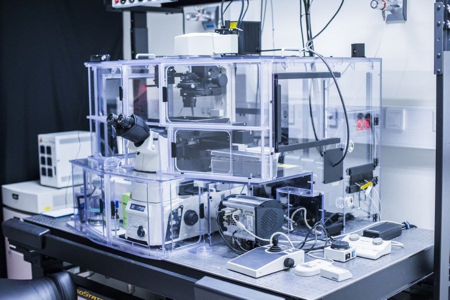
Situated within Tampere University, the Tampere Imaging Facility (TIF) conducts high-quality research and development in bioimaging. TIF provides researchers with state-of-the-art, well-maintained bioimaging equipment and offers training for their use.
Thanks to funding from the European Structural Fund, TIF is embarking on a project to extend its expertise and facilities to local companies and foster the development of novel microscopy technologies. The grant supports a collaborative project between the research groups of Professor Humeyra Caglayan, Professor Atanas Gotchev and Senior Research Fellow and TIF Director Teemu Ihalainen. The three-year project, launched in early 2024, aims to pilot the provision of services to local companies requiring small-scale imaging services.
“Many technology companies stand to gain from our sophisticated equipment and specialised knowledge. Until now, we have lacked standardised processes and staff capacity to handle external tasks or minor projects,” Ihalainen says.
Jenni Karttunen, the appointed Project Manager, is tasked with identifying potential companies that could benefit from TIF’s services and pinpointing the services that are likely to be in demand. The goal is to create an operational model for company collaboration that encompasses the full spectrum from specimen logistics to data reporting. In addition to conducting company pilots, the project participants will develop TIF’s internal processes and author publications on microscopy techniques to provide companies with insights into the potential benefits of partnering with TIF.
“Looking ahead, we hope to expand our offerings to include a wider range of paid services to companies,” Karttunen says.
The ongoing project is laying the groundwork for broader company collaboration.
In addition, the project will pool the expertise of research groups on the Hervanta campus to develop innovative microscopy technologies designed for rapid 3D imaging.
Microscopy images: a fusion of science and aesthetics
In the spring of 2024, TIF organised a microscopy image competition, inviting researchers to submit images taken with TIF’s microscopes. The goal was to highlight TIF’s capabilities while encouraging researchers to appreciate and explore the visual beauty of microscopy images.
“I want to encourage all researchers to not only recognise microscopy images as a rich source of scientific data but also to appreciate their visual and aesthetic qualities,” says Ihalainen.
Visualising these images is important as it enables researchers to emphasise specific aspects of their specimens. For instance, selecting particular colours can greatly enhance the interpretation of the images. An image always conveys more than mere numerical data.
The submissions from the competition will be featured on TIF’s newly launched social media channels on Instagram and X. Through these channels, TIF aims to engage a wider audience with the wonders of science and microscopy.
“People are eager to see beautiful microscopy images. It is an excellent opportunity to simultaneously entertain and educate them about research, cell biology, and microscopy techniques,” Karttunen says.
The winners of the image competition were announced at the Infrastructure Showroom Seminar Series, which was hosted by the Faculty of Medicine and Health Technology and where TIF presented its services to other research centres and researchers at Tampere University. TIF is planning to make the competition an annual tradition. The top three contestants received complimentary microscope time at TIF and a surprise gift.
TIF has a mission to support research
TIF is one of the research infrastructures that are housed at Tampere University to provide researchers with access to cutting-edge technological resources. These research infrastructures encompass an array of specialised instruments and a wealth of expertise, assets that are beyond the reach of individual research groups in terms of acquisition and maintenance.
TIF is equipped with ten light microscopes for examining diverse specimens and one computer dedicated to data analysis. TIF is not only responsible for the upkeep of this sophisticated equipment but also provides training for researchers to ensure they can leverage these tools effectively.
Navigating the delicate balance between high costs and accessibility, TIF is committed to furnishing the research community with high-quality but affordable light microscopy-related imaging services. The support and funding from the Faculty of Medine and Health Technology have a major role in fuelling the continuous evolution and expansion of TIF’s capabilities.


